Jun Lu ![]() ,
You-ting Ju,
Shou-lin Chen,
Jun-ying Cai,
Guo-hai Xu,
Yan-hui Hu
,
You-ting Ju,
Shou-lin Chen,
Jun-ying Cai,
Guo-hai Xu,
Yan-hui Hu
For correspondence:- Jun Lu Email: lujun2611@hotmail.com
Received: 18 January 2016 Accepted: 6 June 2016 Published: 31 July 2016
Citation: Lu J, Ju Y, Chen S, Cai J, Xu G, Hu Y. In situ epicatechin-loaded hydrogel implants for local drug delivery to spinal column for effective management of post-traumatic spinal injuries. Trop J Pharm Res 2016; 15(7):1369-1374 doi: 10.4314/tjpr.v15i7.3
© 2016 The authors.
This is an Open Access article that uses a funding model which does not charge readers or their institutions for access and distributed under the terms of the Creative Commons Attribution License (http://creativecommons.org/licenses/by/4.0) and the Budapest Open Access Initiative (http://www.budapestopenaccessinitiative.org/read), which permit unrestricted use, distribution, and reproduction in any medium, provided the original work is properly credited..
Purpose: To prepare hydrogels loaded with epicatechin, a strong antioxidant, anti-inflammatory, and neuroprotective tea flavonoid, and characterise them in situ as a vehicle for prolonged and safer drug delivery in patients with post-traumatic spinal cord injury.
Methods: Five in situ gel formulations were prepared using chitosan and evaluated in terms of their visual appearance, clarity, pH, viscosity, and in vitro drug release. In vivo anti-inflammatory activity was determined and compared with 2 % piroxicam gel as standard. Motor function activity in a rat model of spinal injury was examined comparatively with i.v. methylprednisolone as standard.
Results: The N-methyl pyrrolidone solution (containing 1 % w/w epicatechin with 2 to 10 % w/w chitosan) of the in situ gel formulation had a uniform pH in the range of 4.01 ± 0.12 to 4.27 ± 0.02. High and uniform drug loading, ranging from 94.48 ± 1.28 to 98.08 ± 1.24 %, and good in vitro drug release (79.48 ± 2.84 to 96.48 ± 1.02 % after 7 days) were achieved. The in situ gel prepared from 1 % epicatechin and 2 % chitosan (E5) showed the greatest in vivo anti-inflammatory activity (60.58 % inhibition of paw oedema in standard carrageenan-induced hind rat paw oedema model, compared with 48.08 % for the standard). The gels showed significant therapeutic effectiveness against post-trauma-induced spinal injury in rats. E5 elicited maximum motor activity (horizontal bar test) in the spinal injury rat model; the rats that received E5 treatment produced an activity score of 3.62 ± 0.02 at the end of 7 days, compared with 5.0 ± 0.20 following treatment with the standard.
Conclusion: In situ epicatechin-loaded gel exhibits significant neuroprotective and anti-inflammatory effects, and therefore can potentially be used for prolonged and safe drug delivery in patients with traumatic spinal cord injury.
Introduction
Flavonoids are the largest group of bioactive polyphenols found in nature. Green and black teas are the richest sources of polyphenols, particularly flavanols and flavonols, which comprise 30 % of the dry weight of the fresh leaf. These dietary flavonoids and nutraceutical compounds—particularly epicatechin (EpC), catechin, and (−)-epigallocatechin-3-gallate (EGCG), which are the chief bioactive constituents—have a broad spectrum of activity. Green tea polyphenols, including EpC, reportedly exert antioxidant, anti-inflammatory, anti-carcinogenic, antiviral, anti-arthritic, antibacterial, antiangiogenic, neuroprotective, and anti-hyperlipidemic effects. These flavones also induce apoptosis and cell cycle arrest in a wide array of cell lines, and protect against cerebral ischaemia [1,2].
Among the various polyphenols, EpC reportedly exhibits a wide range of biological activities, such as antioxidant, antibacterial, anti-carcinogenic, and anti-tumour effects [3,4]. The neuroprotective effects induced by EpC and other various green tea extracts include attenuated ischaemic brain injury and inhibition of excitotoxicity and are evidenced by improved cognitive performance and memory retention; these effects may be utilised in patients with spinal cord injury (SCI) [5–8]. Trauma-induced SCI is one of the most challenging conditions to treat. The primary tissue injury leads to a cascade of adverse events, such as inflammation and apoptosis, which result in neuropathic pain, inflammation, and reversible and/or irreversible damage to the nervous system, known as secondary injury [9–12].
The present work aimed to develop in situ EpC-loaded hydrogels for localised drug delivery to the SC with the advantages of a reduced frequency of dosage, prolonged drug release, and improved patient compliance for the management of SCI. The prepared gels were characterised in terms of their the physicochemical properties, in vitro drug release, in vivo anti-inflammatory activity, and in vivo motor function activity.
Methods
Materials
EpC and chitosan (75 % deacylated, Ch) were obtained from Sigma–Aldrich, US. Other chemicals used were of analytical grade.
Preparation of in situ gels loaded with EpC
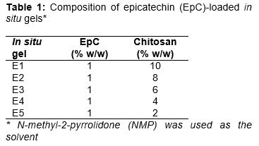
A total of five formulations of pH-responsive in situ gels loaded with EpC were prepared using chitosan (). The polymeric solutions were prepared in NMP. EpC was gently dispersed in the solution with continuous stirring at 200 rpm. The pH of the resulting solution was maintained at 4.0. The solution thus obtained was degassed, followed by membrane filtration sterilisation. The prepared sterile in situ gels were stored in a sterile area in a vacuum desiccator until further use.
Physicochemical characterisation
Clarity was determined by visual inspection against a black and white background. The viscosity of the in situ gelling solution in the presence or absence of simulated biological fluid (SBF) was determined using a Brookfield viscometer. The pH was determined using a digital pH meter.
To determine the drug content of each formulation, each formulation (5 mL removed after vigorous shaking) was mixed with 100 mL of methanol and continuously stirred at 300 rpm for 1 h. The resulting solution was centrifuged at 100 rpm for 10 min. The clear supernatant was then analysed spectrophotometrically to determine the drug content.
In vitro drug release studies
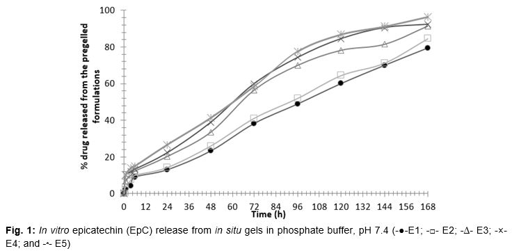
An in vitro drug release study was performed using the method reported by Che et al [13]. The formulations were first brought to a gelled state by mixing 5 mL of the each formulation with 15 mL of phosphate buffer (pH 7.4). This pregelled formulation was stored in a vial containing phosphate buffer (20 mL, pH 7.4) on a shaking water bath (37 ± 1 oC; 50 oscillations per min). Samples (1 mL each) were removed at predetermined time intervals for 1 week; at each sampling time point an equal volume of prewarmed fresh buffer was replaced in the vial. The samples were analysed by ultraviolet (UV) spectrophotometry to determine the drug release at each time point (n = 3).
Animals
Healthy male Wistar rats (150 – 220 g) were used in this study. The rats were kept in cages and housed under standard light and temperature conditions. They were allowed free access to drinking water and a standard diet. The animal study protocols were approved by the Animal Care and Use Committee of Nanchang University, Nanchang (approval no. 2015/02b-13). The studies were carried out in compliance with Directive 2010/63/EU on the handling of animals used for scientific purposes [14].
In vivo anti-inflammatory studies
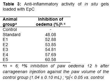
The in vivo anti-inflammatory activity of the formulations was evaluated using the standard carrageenan-induced rat hind paw oedema model. Rats were divided into 6 groups of 6 rats each. The control group (Group I) rats were untreated. For Groups II to V, the in situ gel formulations were injected (20 mg/kg body weight; intra venous) into the left hind paw of the rats. Group VI received the 2 % piroxicam gel (standard). Paw oedema was induced 30 min later by injecting 0.1 mL of a 1 % w/v aqueous suspension of carrageenan into the left paws of all rats. The hind paw volume was measured immediately (0 h) and at predetermined time intervals using a plethysmometer, and was expressed as percent oedema relative to the initial hind paw volume. The percent inhibition of oedema was considered to reflect the anti-inflammatory activity.
In vivo motor function activity
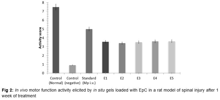
The rats were divided into 7 groups (normal control, negative control, standard, and 4 test groups) of 6 rats each (total, 42 rats). An in vivo motor function activity study was performed as reported by Che et al [13]. With the exception of the animals in the normal control group, all animals were subjected to laminectomy (to induce acute spinal injury) after administration of thiopental sodium (40 mg/kg). Specifically, extradural 40 g force clip compression was performed for 20 s at around T6 (until motor nerve activity is lost). The control group was administered the blank (non-medicated) gel. The test group received the in situ gels (50 mg/kg body weight; intrathecally [i.t.]) at a shaved site closest to the injury. The standard group received intravenous (i.v.) methylprednisolone (30 mg/kg body weight) at 30 min post-injury to allow for drug release. This daily treatment was repeated for 1 week. At the end of 7 days, motor function activity was assessed using a horizontal bar experiment. The time spent by rats on the horizontal bar was determined, and scoring was performed as follows. Activity scores were assigned according to the time spent by each rat on the horizontal bar (1–5 s = 1; 6–10 s = 2; 11–20 s = 3; 21–30 s = 4; >30 s = 5; if bar support touched with forepaw = 5 for 2 mm bar.
Statistical analysis
The results are expressed as means ± standard deviation (SD). Statistical analysis was performed using Origin 9 software (USA) and Student’s t-tests. A value of p < 0.05 was considered to indicate statistical significance.
Results
The gelling capacity of the polymer with SBF was determined, and suitable ratios for maximum gelling were adopted for the formulations.
Physicochemical evaluation
Addition of SBF to the in situ gelling solutions, which were colourless and clear when inspected visually, resulted in formation of a viscous gel mass. The in situ gel formulation solution showed a uniform pH in the range of 4.01 ± 0.12 to 4.27 ± 0.02 ().
The formulation showed high and uniform drug loading, ranging from 94.48 ± 1.28 to 98.08 ± 1.24 %. The viscosity of the formulations was found to increase 5- to 7-fold () after gelling. The formulations showed a shear-thinning property (), as has been reported previously.
In vitro drug release studies
In vitro drug release studies were performed in phosphate buffer at pH 7.4 to simulate the pH of biological fluid. The formulations showed drug release proportions ranging from 79.48 ± 2.02 to 96.48 ± 1.05 % after 7 days ().
In vivo anti-inflammatory activity
Inhibition of carrageenan-induced paw oedema was taken as evidence of anti-inflammatory activity (). The reduction in the initial volume of paw oedema over time due to the effect of the standard drug or test formulation was assessed. The percent inhibition of paw oedema ranged from 52.88 to 60.58%, compared with 48.08 % for the standard.
In vivo motor function activity
The motor nerve activity elicited by the formulations was assessed using a rat model of post-traumatic SCI. After treatment for 7 days, the activity scores of the rodents treated with the in situ gels were 3.56, 3.4, 3.5, 3.6, and 3.62 for E1, E2, E3, E4, and E5, respectively. E5 elicited the optimum motor nerve activity in rats with traumatic SCI ().
Discussion
Each year, several million people suffer from serious traumatic peripheral nerve injuries globally. If the damaged nerves cannot be restored, loss of muscle function, impaired sensation, and painful neuropathies can result. Acute steroids have been reported to be an effective first-line therapy in traumatic SCI; indeed, a high dose of methylprednisolone given immediately after traumatic SCI is an accepted standard treatment [15]. Despite several reports of the effectiveness of i.v. methylprednisolone in traumatic SCI-induced nerve damage, it cannot cross the blood–spinal cord barrier. This results in the need for frequent administration of large doses of methylprednisolone.
Biodegradable polymers in the form of scaffolds, implants, or hydrogels are safe and effective platforms for site-specific or targeted drug delivery [16–19]. Localised drug delivery via intrathecal injections of polymeric systems in the SC likely elicits results comparable to those of conventional parenteral steroid therapy, but with a lower dose and prolonged drug release [13,20,21]. Injectable in situ hydrogel implants have been investigated in terms of their neuroprotective effects against SCI. These polymeric solutions are injectable and are converted into a hydrogel (i.e., viscous matrix depot form) due to a change in temperature, pH, ion cross-linking, or solvent removal [22–24].
In the present study, we prepared chitosan-based in situ hydrogel implants; hydrogel formation was triggered by a change in pH and solvent removal [25]. Immediately after contact with an aqueous environment, the NMP solvent diffuses out, resulting in polymer precipitation. This ultimately results in the formation of a porous polymer matrix, which slowly releases the drug into the biological microenvironment. High drug loading was observed with the in situ gels showed, a property desirable for drug delivery. The rheology of the prepared formulation was also satisfactory with the consistency in which it could be easily injected. The viscosity of the formulation was governed by the nature and amount of polymer present. In principle, the in situ gel should not alter the basic rheological properties of the biological fluid at the site of action. In this case, the formulation was delivered into the intrathecal fluid, which is crucial for neuroprotection. Further studies to assess the effect of the in situ formed hydrogel on the rheology of the intrathecal fluid are warranted.
The formulation with low chitosan content had better release properties. Use of a higher polymer content might have resulted in the formation of a stiffer gel, which would act as a barrier to drug diffusion. The in situ gel formulation E5 showed the highest anti-inflammatory activity, 60.58 % inhibition of paw oedema compared with 48.08 % for the standard. This might have been due to the lower polymer concentration, which facilitated drug release. However, the E1, E2, and E3 formulations may function better over time, as these released the drug over a prolonged period of time. Moreover, all of the formulations had better results than those seen following treatment with the standard.
The horizontal bar method is a practical and easy-to-use method of assessing motor nerve activity in rats with post-traumatic SCI. The activity score of the model rats following treatment with the formulations was 3.4 – 3.6, and that following treatment with the standard was 5.0. Therefore, the motor nerve activity was greater after treatment with the EpC-loaded in situ gels than that following treatment with the standard.
Conclusion
Biodegradable polymers can be used in in situ EpC-loaded gels for continuous, extended drug release for a round-the-clock management of pain and inflammation in patients with SCI. The gel prepared from EpC and chitosan at a 1:4 ratio exhibited the greatest potential for prolonged post-traumatic therapy of SCI due to its considerable neuroprotective and anti-inflammatory effects.
References
Archives


News Updates